All issues
Author:Hui-Fang Ni*, Ching-Yi Lin, and Chao-Jung Wu
Abstract:
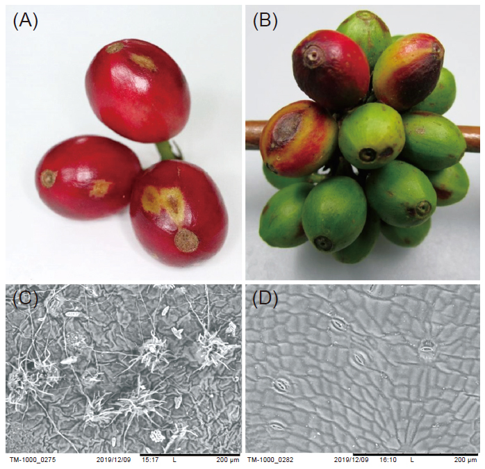
Coffee brown eye spot disease, caused by Cercospora coffeicola, mainly occurred between September and December and reached its peak period in November. In the initial stage, needle spots with ashy centers appeared on coffee leaves. The spots expanded with gradually visible yellow halo. On berries, ashy oval spots grew and became brown lesions, and some lesions had bright purple halo. The pathogen produced aggregated conidiophores both on upper and lower sides of coffee leaves. The aggregated conidiophores were produced from stomata on lower side of leaves. On upper side, conidiophores appeared on the lesions. The pathogen grows in the range of 20–30℃, and 25℃ is the optimal growth temperature. After 6 h of incubation in 20–35℃, the spore germination ratio was 90%. With free water at 25℃, the spore germination ratio reached 60% in 3 h. Spore germination was inhibited under 10℃ and above 40℃. No wound was needed when the pathogen invaded from the lower sides of leaves. However, wounding was necessary for infection on the upper side of leaves, and lenticels on berries might be a possible path for invasion. The pathogen was hardly sporulated on potato dextrose agar (PDA), and the colony was gray-black and grows slow. Some isolates secreted red pigment on PDA. Using recommended fungicides and fungicides with potential to become extended recommended chemicals, the fungicide screening tests were conducted. The results showed that, among the recommended fungicides, Pyraclostrobin inhibited mycelial growth and spore germination at the same time. Instead, Tebuconazole inhibited mycelial growth only. For fungicides with potential to become extended recommended chemicals, Fluazinam inhibited both mycelial growth and spore germination, and cyprodinil + fludioxinil was only effective on mycelial growth. The above results might provide the information for controlling coffee brown eye spot disease.
Key words:Coffee, Coffee brown eye spot disease, Fungicide screening
Download:![]() PDF Links
PDF Links
Taiwan Agricultural Research
 Submit your manuscript
 Guide for authors
- 1. Using Digital Soil Mapping to Predict Soil Organic Carbon Stocks in Zhuoshui River Basin
- 2. Development of a Technique for Forecasting (or Pre-Detection) Anthracnose Disease Incidences of Green Mature Bagging Mango Fruits
- 3. Taxonomic Review of the Genus Asiophrida Medvedev, 1999 in Taiwan (Insecta: Coleoptera: Chrysomelidae: Galerucinae: Alticini), with Notes on Biology